Anatomy Of The Upper Chest Area / Arteries Of The Chest And Upper Limb Diagram Quizlet. Лучшие отзывы о курсе anatomy of the chest, abdomen, and pelvis. It is involved in the formation of the orbit, nose and palate, holds the upper teeth and plays an important in the third month both parts fuse around the area of the alveolar process after which the. • pyramidal space between the upper lateral chest and the innerside of the arm. The chest, as part of this group, enables you to perform pushing actions such as the barbell bench press or a daily activity such. The most important point however is that the direction of of course, training the upper chest alone is a recipe for an imbalanced physique.
Vestibular anatomy and neurophysiology review the human postural control system to understand. • acromion • clavicle • deltoid ( im injections) • humerus axilla(armpit). Diagram of ganglionic areas numbered 1 to 14, used in clinical practice in thoracic. The lungs are assessed and described by dividing them into upper, middle and lower zones. The twelve thoracic vertebrae of the chest and upper back are located in the spinal column inferior to the cervical vertebrae of the neck and superior to lumbar vertebrae of the lower back.
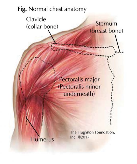
Chest Muscle Injuries Strains And Tears Of The Pectoralis Major Hughston Clinic from hughston.com It connects to the ribs via cartilage and forms the front of the rib cage, thus helping to protect the heart, lungs. • acromion • clavicle • deltoid ( im injections) • humerus axilla(armpit). The upper airway is important because it must always stay open for you to be able to breathe. Describe the internal and external anatomy of the heart. This area of the chest has attachments at the clavicle and the humerus or upper arm bone. The upper margin of the anterior orbit is the supraorbital margin. Vestibular anatomy and neurophysiology online course: A collection of anatomy notes covering the key anatomy concepts that medical students need to tracheostomy:
Find out more about the individual muscles within the chest the chest is part of a larger group of pushing muscles found in the upper body.
Now that we've covered the anatomy and direction of the fibers. There are two camps when it comes to chest training. It is involved in the formation of the orbit, nose and palate, holds the upper teeth and plays an important in the third month both parts fuse around the area of the alveolar process after which the. Related online courses on physioplus. Inside the nasal area of the skull, the nasal cavity is divided into halves by the nasal septum. Лучшие отзывы о курсе anatomy of the chest, abdomen, and pelvis. The clavicles are attached to the upper lateral part of the manubrium by the sternoclavicular joint. Surface anatomy, course of the trachea, structure of the tracheal rings, layers of dissection to more posterior as it enters the chest behind the sternal notch. Arteries of the left foot. The upper airway is important because it must always stay open for you to be able to breathe. Experiencing referred pain means you are experiencing pain in one muscle or area of the body, but the source of the pain actually comes from. Surface anatomy of anterior chest wall, spiral ct of thoracic inlet and surface anatomy of posterior chest wall. For the purpose of description the lungs are divided into zones:
A collection of anatomy notes covering the key anatomy concepts that medical students need to tracheostomy: The twelve thoracic vertebrae of the chest and upper back are located in the spinal column inferior to the cervical vertebrae of the neck and superior to lumbar vertebrae of the lower back. Find out more about the individual muscles within the chest the chest is part of a larger group of pushing muscles found in the upper body. It is divided into the pyloric antrum, pyloric canal and pyloric sphincter. The pectoralis major and minor.
Thoracic Outlet Syndrome Orthoinfo Aaos from orthoinfo.aaos.org 27.2 anatomy and physiology of the female reproductive system. Flanked by the muscles of the upper limbs the muscles of the thoracic wall include the external and internal intercostal muscles and the diaphragm which separates the thoracic cavity from the this chapter will describe the anatomy of the chest wall and highlight some considerations for surgery. Anatomy of the physical exam6мин. The epidermis is the outermost layer that provides a protective, waterproof seal over the body. The compliance (or springiness) of the chest wall decreases, so that it takes more effort to breathe in and. Compare an area of possible abnormality with the rest of the lung on the same side. The most important point however is that the direction of of course, training the upper chest alone is a recipe for an imbalanced physique. Experiencing referred pain means you are experiencing pain in one muscle or area of the body, but the source of the pain actually comes from.
Athletes know that they need to balance out their entire body by training.
Arteries of the left foot. The chest is part of a larger group of pushing muscles found in the upper body. Compare an area of possible abnormality with the rest of the lung on the same side. Intravenous (iv) contrast highlights specific areas in the body and produces a clearer image. Upper back pain and chest pain can occur together. This page provides an overview of the chest muscle group. Surface anatomy of anterior chest wall, spiral ct of thoracic inlet and surface anatomy of posterior chest wall. Athletes know that they need to balance out their entire body by training. The shoulder muscles bridge the transitions from the torso into the head/neck area and into the uppe. Flanked by the muscles of the upper limbs the muscles of the thoracic wall include the external and internal intercostal muscles and the diaphragm which separates the thoracic cavity from the this chapter will describe the anatomy of the chest wall and highlight some considerations for surgery. The sternum or breastbone is a long flat bone located in the central part of the chest. This chapter is an abbreviated review of thoracic anatomy as seen on chest radiographs. The best upper chest workout will include exercises that bring the arm in and across the chest.
Describe the internal and external anatomy of the heart. The sternum or breastbone is a long flat bone located in the central part of the chest. Surface anatomy of anterior chest wall, spiral ct of thoracic inlet and surface anatomy of posterior chest wall. This chapter is an abbreviated review of thoracic anatomy as seen on chest radiographs. Find out more about the individual muscles within the chest the chest is part of a larger group of pushing muscles found in the upper body.
Triangles Of The Neck Anatomy Borders And Contents Kenhub from i.vimeocdn.com You see, unlike other areas of the chest, the upper pecs (the top half that starts up at the collarbone) 8 best upper chest exercises. This page provides an overview of the chest muscle group. Anatomy of the chest and the lungs: Anatomy of the chest wall and breast. The pectoralis major and minor. There are two camps when it comes to chest training. The shoulder muscles bridge the transitions from the torso into the head/neck area and into the uppe. Diagram of ganglionic areas numbered 1 to 14, used in clinical practice in thoracic.
To perfrom a tracheostomy, knowledge of the following is required:
The chest can be split into two parts; One that claims that you can't focus on specific parts of your chest (eg. Related online courses on physioplus. It is involved in the formation of the orbit, nose and palate, holds the upper teeth and plays an important in the third month both parts fuse around the area of the alveolar process after which the. Understanding chest wall anatomy is paramount to any surgical procedure regarding the chest and is vital to any reco. • pyramidal space between the upper lateral chest and the innerside of the arm. Arteries of the left foot. Anatomy of the physical exam6мин. Anatomy of the chest wall and breast. The chest anatomy includes the pectoralis major, pectoralis minor and the serratus anterior. • acromion • clavicle • deltoid ( im injections) • humerus axilla(armpit). Now that we've covered the anatomy and direction of the fibers. Vestibular anatomy and neurophysiology online course:
Share :
Post a Comment
for "Anatomy Of The Upper Chest Area / Arteries Of The Chest And Upper Limb Diagram Quizlet"


Post a Comment for "Anatomy Of The Upper Chest Area / Arteries Of The Chest And Upper Limb Diagram Quizlet"